On 31 July 2025, a special-purpose subsidy project entitled “Infrastructure for high‑throughput quantitative and qualitative analysis of pico‑ and nanoplankton” was completed. The project leader was Dr. Katarzyna Piwosz, Professor of NMFRI, from the Department of Fisheries Oceanography and Marine Ecology.
Within the scope of the project, the following equipment was purchased:
- A ZE5 flow cytometer by Bio‑Rad with an automatic sample loader, costing PLN 1,296,954.90.
- An automated Nikon ECLIPSE NI‑E epifluorescence microscope with digital image analysis software, costing PLN 619,999.92.
About the Flow Cytometer
It is equipped with five lasers (375 nm, 405 nm, 488 nm, 561 nm, and 640 nm), 27 fluorescence detectors, and three light scatter detectors (including a small-particle detector at the 405 nm laser), enabling simultaneous analysis of up to 30 populations based on fluorochrome staining and natural pigment autofluorescence (chlorophylls, phycocyanins, phycoerythrins). The automatic sample loader reduces manual involvement, allowing up to 300 samples to be processed in an 8-hour workday, significantly increasing throughput and research efficiency. The cytometer is used for quantitative studies of viruses, prokaryotes (bacteria and archaea), and picophytoplankton.
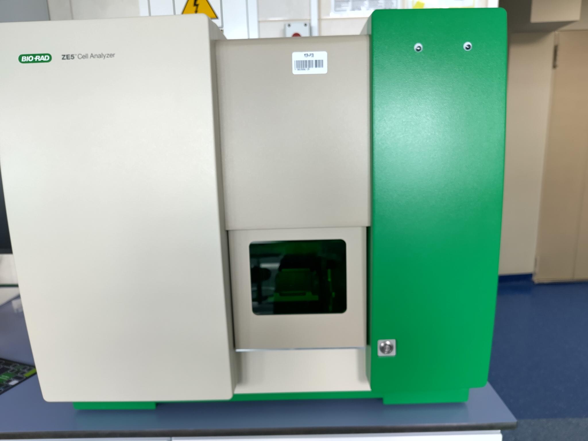
Fig. 1. ZE5 flow cytometer by Bio-Rad
About the Automated Epifluorescence Microscope
The motorized Nikon ECLIPSE NI‑E microscope includes a scanning stage for eight microscope slides, image acquisition and analysis software utilizing machine learning algorithms, seven LED light sources (385 nm, 430 nm, 475 nm, 555 nm, 590 nm, 630 nm, 735 nm), nine filter blocks, objectives ranging from 1× to 100× magnification, and a camera with 5000 × 2900 pixel resolution. Automation accelerates traditionally labor-intensive microscopy workflows, greatly improving analysis throughput and precision by reducing human error. It is used for quantitative and qualitative analysis of prokaryotic communities, pico- and nanophytoplankton, pico- and nanoplanktonic protozoa using methods such as CARD‑FISH, and for studying feeding interactions between protozoa and bacteria by observing food vacuoles.
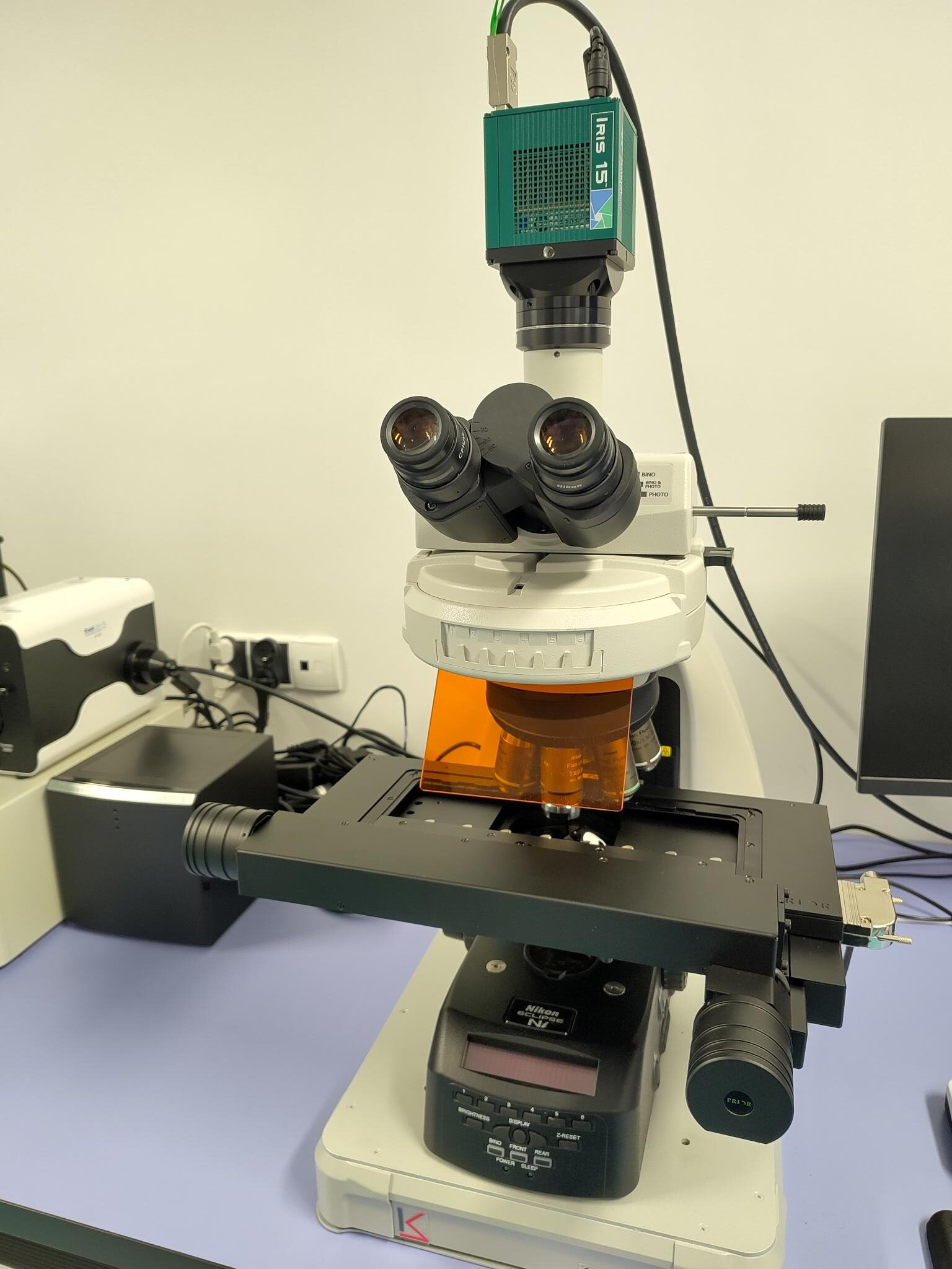
Fig. 2. ECLIPSE NI-E automated epifluorescence microscope by Nikon
The new infrastructure supports innovative research avenues aligned with the EU Strategy for the Baltic Sea Region under the thematic pillar “Sea protection”, contributing to the following specific objectives:
- Clear water in the sea
- Rich and healthy wildlife
- Clean and safe shipping
These capabilities enable focused applications such as monitoring toxic algal blooms (e.g., Prymnesium parvum), tracking the ecological status of the Baltic Sea at the microbial level (bacteria, pico‑ and nanoplankton), and studying the impact of pollutants on microorganisms.
Lastly, the new infrastructure is available for collaborative use by other institutions through partnerships with the National Maine Fisheries Research Institute.

Special-purpose subsidy from the Ministry of Science and Higher Education